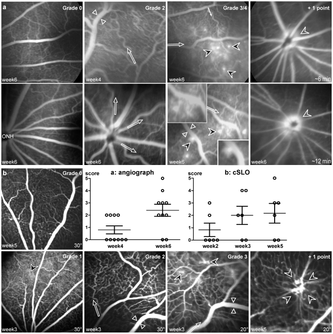
Figure 2. Fluorescein angiograms of Brown-Norway rat eyes.
(a) Observations with a classic angiograph (Pro III Fundus camera, Kowa), 4 and 6 weeks after pVAX2-rPGF ET. Angiograms were established with a scan angle of 30°. Vascular abnormalities were scored from 0 to 5 in accordance to the following grading: Grade 0, normal retinal vasculature, as observed in control fundus at week 6 (ONH, Optic Nerve Head); +1 point for each of the following changes - dilated (between white arrowheads) or tortuous (white arrows) vessels, microaneurysmal-like hyperfluorescent dots (black arrowheads) <10 or hyper-fluorescence around the ONH; +2 points for microaneurysmal-like hyper-fluorescent dots >10. (b) Observations with a confocal scanning Laser Ophthalmoscope (cSLO, Heidelberg Retina Angiograph I), 2, 3 and 5 weeks after pVAX2-rPGF ET. The same grading was used to score vascular abnormalities. The higher resolution of the cSLO allowed the observation of early fluorescein leakage (Grade 1), and the detection of strong vascular abnormalities at later stage (+ 1 point).

