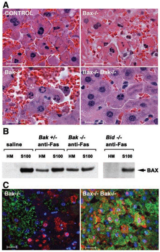
Fig. 3.
Function of BAX and BAK downstream of BID in Fas-induced hepatocellular apoptosis. (A) Hematoxylin and eosin-stained liver sections from Bak−/−, Bax−/−, and DKO mice treated with antibody to Fas (anti-Fas). Bar represents 20 µm. (B) Fas-induced BAX translocation from cytosol to mitochondria in hepatocytes is downstream of BID but independent of BAK. BAX immunoblot of mitochondrial (HM) and cytosolic (S100) fractions of hepatocytes prepared as previously described (8) from wild-type, Bid−/−, Bak+/−, and Bak−/− mice treated with saline or anti-Fas. (C) Three-color immunohistochemistry of livers from anti-Fas–treated Bak−/− and DKO mice. Cytochrome c staining is in green, activated Caspase-3/7 (CM1 antibody) in red, and nuclei stained with Hoechst in blue. Bar represents 20 µm. Histology and immunohistochemical staining of livers from anti-Fas–treated animals were done as described (14).

