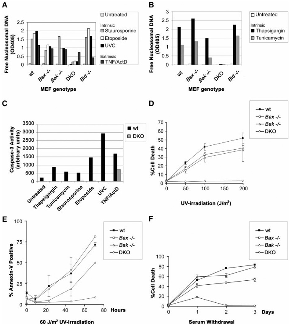
Fig. 4.
Resistance of Bax, Bak doubly deficient MEFs to multiple intrinsic death signals. (A) Susceptibility of MEFs to apoptotic death by mitochondria-dependent intrinsic signals. Wild-type, Bax−/−, Bak−/−, DKO, and Bid−/− primary MEFs were treated with 1 µM staurosporine, 100 µM etoposide, UVC (60 J/m2), or TNF-α (1 ng/ml) + actinomycin D (2 µg/ml), and a 48-hour time point is shown. Average values from duplicate samples of an enzyme-linked immunosorbent assay of apoptotic DNA fragmentation (Roche) are plotted as representative of three independent experiments. (B) Susceptibility of MEFs to apoptotic death by ER stimuli. As in (A), genotyped MEFs were treated with 2 µM thapsigargin or tunicamycin (1 µg/ml), and average values from duplicate samples at a 48-hour time point of apoptotic DNA fragmentation were plotted. DKO MEFs also demonstrated long-term survival when assessed by Annexin-V staining 4 days after the stimuli (12). (C) Quantitation of effector caspase activity (e.g., Caspase-3) using DEVD-AFC fluorogenic substrate (Clontech). Wild-type and DKO MEFs were treated with the following death signals and harvested at indicated time points: 2 µM thapsigargin (36 hours), tunicamycin (1 µg/ml; 36 hours), 1 µM staurosporine (16 hours), 100 µM etoposide (24 hours), UVC (60 J/m2; 24 hours), and TNF-α (1 ng/ml) + actinomycin D (2 µg/ml; 16 hours). Results are representative of three independent experiments. (D) Dose response of MEFs to UVC irradiation. Cell death was quantitated by trypan blue exclusion, and a 24-hour time point was plotted. (E) Time course of MEF apoptosis after exposure to UVC (60 J/m2). Cell death was quantitated by flow cytometric detection of Annexin-V staining. Long-term survival of DKO MEFs was also noted 4 days after staurosporine and etoposide, as well as UVC treatments (12). (F) Time course of MEF apoptosis in response to serum withdrawal. Cell death was quantitated by trypan blue exclusion.

