
Fig. 3.
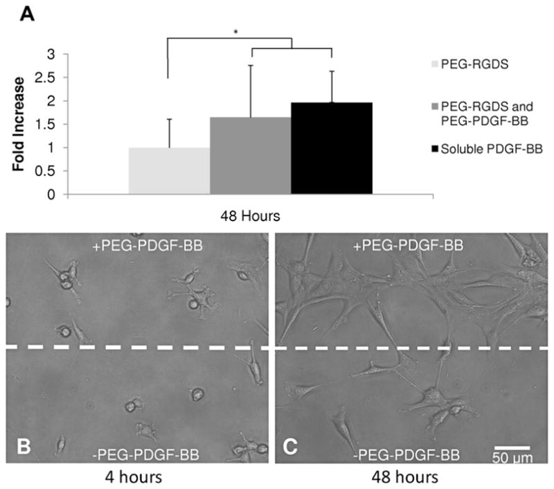
Bioactivity of PEG–PDGF was confirmed by seeding 10T1/2 cells onto modified surfaces. (A) By 48 h 10T1/2 cell proliferation had increased significantly in the presence of PDGF-BB (ANOVA, #P < 0.05). No significant difference between the soluble and bound forms of PDGF-BB indicate that covalently immobilized PEG–PDGF-BB retained its bioactivity. (B, C) Hydrogels with patterned regions of PEG–PDGF-BB demonstrate the utility of spatial control of PDGF-BB. 10T1/2 cells seeded onto these patterned gels were evenly dispersed at 4 h (B), but exhibited increased proliferation on regions patterned with PDGF-BB by 48 h (C).