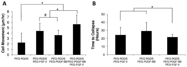
Fig. 7.
(A) HUVECs encapsulated in 3D degradable hydrogels exhibited greater cell movement in gels containing covalently immobilized growth factors (*P < 0.01). PEG–FGF-2 significantly increased migration compared with PDGF-BB (#P < 0.05), and the combination of PEG–PDGF-BB and PEG–FGF-2 induced significantly increased cell migration compared with PDGF-BB and FGF-2 alone (*P < 0.01). (B) Hydrogels containing covalently immobilized growth factors collapsed after 20–40 h. The combination of PDGF-BB and FGF-2 led to earlier gel collapse compared with each growth factor alone (*P < 0.01).

