
FIG. 2.
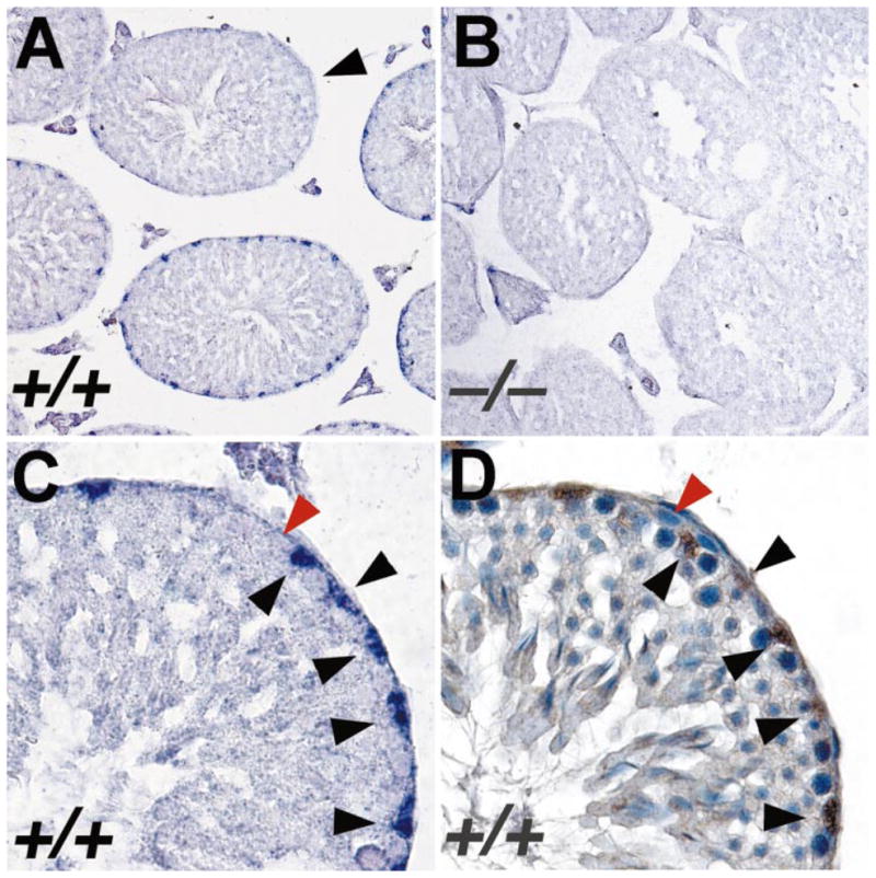
In situ hybridization analysis of expression of Bclw mRNA in mouse testis. A) Section of testis from adult wild-type mouse hybridized with an antisense ribo-probe derived from the 3′-UTR of Bclw cDNA. A significant positive signal (a purple precipitate) is observed only over nuclei of Sertoli cells. Significant signal was observed in all stages of tubules, except those between stages VII and VIII of the seminiferous cycle (arrowhead). ×200. B) Section of testis from adult Bclw homozygous mutant prepared and analyzed simultaneously under identical conditions as those in A. Only background purple precipitate is seen. ×200. C) Higher magnification of a single seminiferous tubule from a wild-type mouse, hybridized with anti-sense Bclw 3′-UTR riboprobe. Sertoli cell nuclei (black arrowheads) stain relatively intensely purple. In contrast, a spermatogonium (red arrowhead) does not display any significant staining over background. ×640. D) Serial section from that shown in C reacted with antibody for GATA-1, a marker of Sertoli cell nuclei. The brown staining denotes presence of the antibody. The blue counterstain is hematoxylin. Black and red arrowheads denote the same cells as identified in C. ×640.