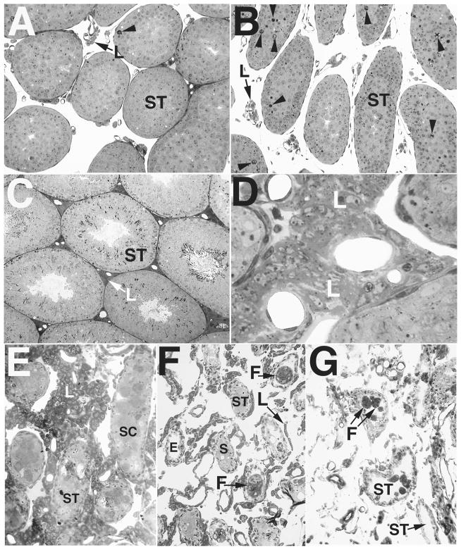
FIG. 4.
Light micrographs showing development of the mutant testicular phenotype in Bclw-deficient mice, emphasizing the general appearance of the testis and the Leydig cells. A and B) Testes at 20 days of age showing the similar appearance of control (A) and Bclw-mutant animals (B) with respect to Leydig cells (L). As previously described [5], approximately sevenfold more degenerating cells (arrowheads) are seen in the seminiferous tubules (ST) from mutants than in the control. ×70 and ×60, respectively. C and D) Testes from 3-mo-old animals showing development of the Leydig cells (L) in control (C) and Bclw-mutant animals (D). Intertubular clusters of Leydig cells are extensive in Bclw mutants. ×80 and ×350, respectively. E) Testes from 5.5-mo-old mutants showing the prominent presence of Leydig cells (L). Large masses in seminiferous tubules (ST) were composed of sloughed dead and or dying Sertoli cells (SC). ×155. F) Testes from 8-mo-old mutants showing small “strings” of Leydig cells (L) among extremely small seminiferous tubules (ST), with the latter containing some Sertoli cells (S) or foreign-body giant cells (F). A seminiferous tubules devoid of cellular contents (peritubular “ghosts”) is marked (E). ×75. G) Testes from 12-mo-old mutants with absence of Leydig cells. The scattered cells between peritubular “ghosts” are connective tissue elements and phagocytic elements. A few foreign-body giant cells (F) are seen within seminiferous tubules (ST). ×100.

