Fig. 6.
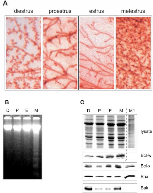
Bak is implicated as a cell-death gene during mammary epithelial apoptosis in the estrous cycle. (A) Whole mounts of mammary gland of ICR mice at different times of the estrous cycle stained with Haematoxylin show the ductal structures in estrus, the extent of proliferation within 2 days at metestrus, and the progressive regression of the next few days through diestrus and proestrus. (B) 5 μg genomic DNA extracted from mammary gland from the same mice was subjected to agarose gel electrophoresis. Apoptotic DNA ladders appear during metestrus (M) and are absent in proestrus (P) and estrus (E). We did not detect ladders in the diestrus (D) sample by this method, possibly because of its lack of sensitivity, although apoptosis has previously been shown to occur at this stage of the cycle by in situ methods (Andres et al., 1995). (C) Whole-tissue lysates were prepared from mammary gland at different times of the estrous cycle and subjected to polyacrylamide gel electrophoresis. Coomassie staining indicates that the levels of protein were even in each sample (top panel). Similar gels were transferred to nylon and immunoblotted using antibodies directed against Bcl-w, Bcl-x, Bax and Bak. The levels of Bak in estrus correspond to those in the virgin tissue extracts shown in Fig. 2F, whereas its increased levels at metestrus correlate with the induction of apoptosis. Bad could not be detected in non-pregnant tissue at any stage of the estrous cycle.

