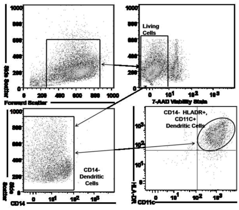
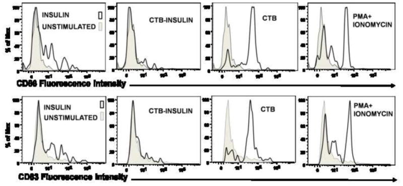
Figure 3. Dendritic Cell Activation and Maturation is Suppressed by CTB-INS Fusion Protein.


(Panel A) Flow cytometry dot plot showing dendritic cells differentiated from monocytes isolated by ficoll gradient centrifugation from human umbilical cord blood. The CD14+ monocytes were supplemented with granulocyte macrophage colony stimulating factor (GMCSF) and IL-4 and were cultured for 6 days to obtain CD14-HLA-DR+CD11c+ immature human DCs. To determine the viability of DCs isolated from the entire population of collected cells (Upper left panel), 7-AAD negative cells were gated (Upper right panel) and analyzed for expression of the surface marker CD14 (Lower left panel). The CD14- cells were gated and analyzed for co-expression of HLA-DR and CD11C differentiation markers by flow cytometry. (Panel B) The histograms depict the expression of dendritic cell CD83 and CD86 after stimulation of DCs for 48 hours with CTB, CTB-INS, Insulin and PMA Ionomycin. Maturation state of the DCs was determined by flow cytometric analysis. The data are representative of three independent experiments with comparable results.
