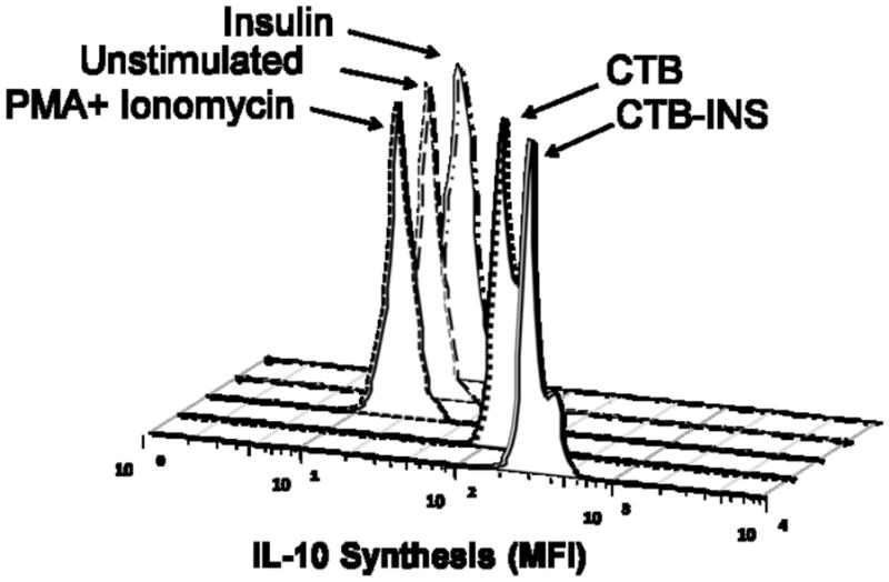
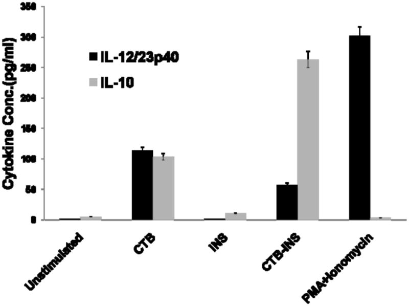
Figure 6. CTB-INS Fusion Protein Stimulates IL-10 Synthesis.


(Panel A) The histograms show the mean fluorescence intensity of IL-10. Premixed plastic beads coated with capture antibodies (BD Biosciences, San Jose, CA, USA).) and a mixture of Phycoerythrin-conjugated antibodies against IL-10 were incubated for 2 hours with the supernatant removed from different treatment conditions of proteins with DCs as described in the Materials and methods section. The beads were washed and analyzed by flow cytometry for determination of the fluorescence intensity of bound IL-10. (Panel B) shows Cytometric Bead Array-defined concentrations of IL-10 and IL-12/23p40 subunit measured from cell supernatants taken from DCs receiving different treatment conditions. For each treatment the concentration of IL-10 and IL-12/23p40 subunit was normalized to standard IL-10 and IL-12/23p40 cytokine curve and given in pg/ml. The data are the Mean and SE (*P<0.001) of repeated independent experiments as compared to the control sample.
