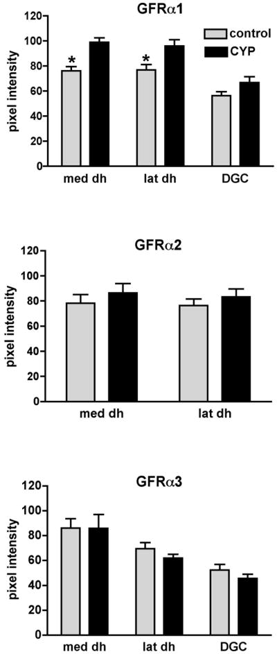
Figure 7.
Densitometric analysis of GFR-immunoreactivity in sacral spinal cord of control and cyclophosphamide (CYP)-treated animals. Histograms show mean ± SE of raw pixel intensity measurements taken from 6–10 randomly selected sections for each group and immunostain. CYP treatment caused a significant increase in GFRα1-IR pixel intensity in both medial and lateral dorsal dorsal horn but had no significant effect on the dorsal gray commissure, although there appeared to be a small increase (medial, P=0.002, lateral, P= 0.02, DGC, P=0.08, unpaired t test; n=6 control, n=4 CYP). CYP treatment had no effect on GFRα2- or GFRα3-IR pixel intensity (n=4 for both control and CYP groups). med dh, medial dorsal horn; lat dh, lateral dorsal horn; DGC, dorsal grey commissure.

