Figure 2.
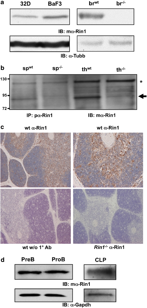
Endogenous mouse Rin1 expression. (a) Left: Immunoblot (IB) of Rin1 in 32D and BaF3 cells. Right: IB of wild-type (wt) and Rin1−/− (−/−) brain (br) tissue confirming antibody specificity. β-tubulin (Tubb) control below each blot. (b) IB of immunoprecipitated (IP) material from spleen (sp) and thymus (th) tissue of wt and Rin1−/− mice. Arrow indicates Rin1. Asterisk marks background band. mα, monoclonal antibody; pα, polyclonal antibody. (c) Immunohistochemical stain of Rin1 in mouse thymus. Top two panels show wild-type thymus probed with anti-Rin1 and counter stained with hematoxylin (left, × 4; right, × 10). Bottom left panel shows wild-type control without anti-Rin1. Bottom right panel shows Rin1−/− thymus control. (d) IB of Rin1 in PreB, ProB and common lymphoid progenitor (CLP) cells. Gapdh control below.

