Figure 5.
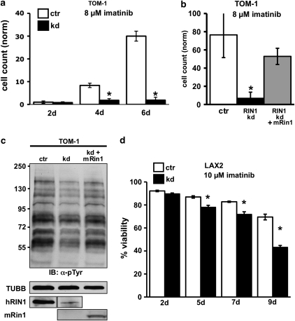
RIN1 silencing sensitizes ALL cells to imatinib. (a) Control (ctr) and RIN1-silenced (kd) TOM-1 cells (1 × 10⁴/ml) were cultured in 8 μ imatinib for the indicated time. Cell counts normalized to 2d-ctr. (b) Control (ctr), RIN1 knockdown (RIN1 kd) and knockdown rescued with mouse Rin1 (RIN1 kd+mRin1) TOM-1 cells were cultured in 8 μ imatinib for 9 days. (c) Control, RIN1 kd and RIN1 kd+mRin1 TOM-1 cells were immunoblotted with anti-phosphotyrosine. TUBB and RIN1 immunoblots are shown below. Murine Rin1 and human RIN1 were detected using different antibodies. Note: hRIN1 bands are from the same exposure of a single immunoblot. (d) Control (ctr) and RIN1-silenced (kd) B-ALL cells were cultured in 10 μ imatinib for the indicated time. Cell viability was determined by propidium iodide stain and flow cytometry. Panels a, b and d: s.d. from triplicate samples counted in duplicate; * indicates P<0.05 between control and knockdown.

