
Figure 4. Marked gain-of-function for Kir6.1-S422L channels.
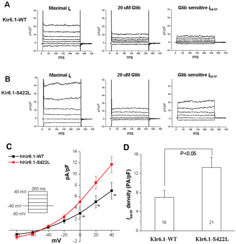
Comparing the glibenclamide (Glib)-sensitive KATP current of Kir6.1-S422L with Kir6.1-WT. (A) Examples of whole cell current traces of Kir6.1-WT recorded after 1-2 minutes perfusion of 100 μM pinacidil (maximal Ik, left), in the presence of 20 μM Glib (middle), and the Glib-sensitive IKATP obtained by subtraction (right). (B) Examples of whole cell current traces of Kir6.1-S422L recorded after 1-2 minutes perfusion of 100 μM pinacidil (maximal Ik, left), in the presence of 20 μM Glib (middle), and the Glib-sensitive IKATP obtained by subtraction (right). (C) Summary data of the current voltage plot for the Glib-sensitive IKATP at the voltages tested (insert, n=16-21 cells. * p<0.05). (D) Bar graphs showing the mean IKATP densities from Kir6.1-WT and Kir6.1-S422L channels elicited by a test pulse from holding potential of -40 mV to 40 mV for 260 ms. IKATP density of Kir6.1-S422L mutant channels was increased significantly compared with Kir6.1-WT channels.