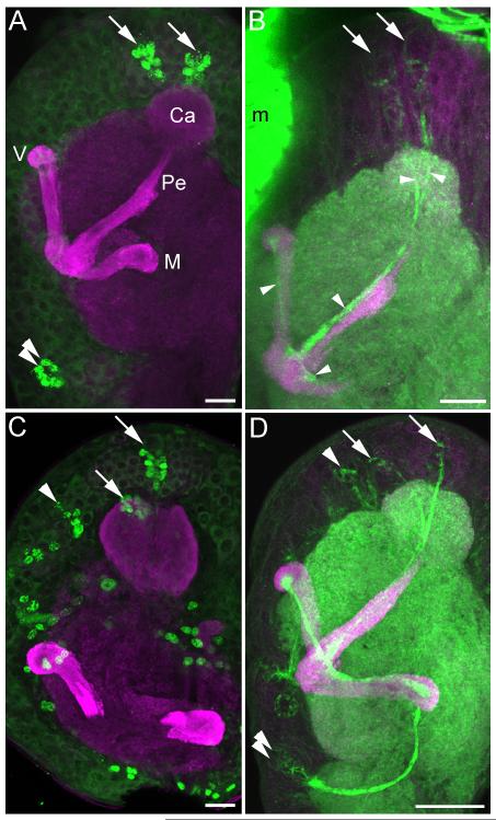
Figure 2. Mushroom body development and neurogenesis in first and second instar larvae.
Anti-DC0 (magenta, all panels) staining shows simple mushroom bodies in the Manduca sexta larva, composed of a single calyx (Ca), medial lobe (M), vertical lobe (V) and pedunculus (Pe). A. BrdU labeling (green) in the first instar reveals neurogenesis by only three neuroblasts per hemisphere, the two mushroom body neuroblasts (arrows) and one neuroblast in the deutocerebrum (double arrows). B. Phalloidin labeling (green) is concentrated in the membranes of proliferating neuroblasts, newborn Kenyon cells beneath each mushroom body neuroblast (arrows) and their axons extending through the calyces and into the pedunculus and lobes (arrowheads). C. BrdU labeling (green) in the second instar larva reveals additional neurogenesis throughout the brain, including the mushroom body neuroblasts (arrows) and a protocerebral neuroblast anterior to the calyx (arrowhead). Tracing this latter neuroblast through larval development suggests that it contributes to the Class III Kenyon cell pool (see Figure 3). D. Phalloidin labeling (green) reveals newborn Kenyon cells produced by the mushroom body neuroblasts (arrows) and likely Class III Kenyon cells produced by the anterior protocerebral neuroblast (arrowhead). Axons of newborn neurons generated by the deutocerebral neuroblast are also labeled with phalloidin (double arrowheads). m- muscle, Scale bars = 20 μm for all panels.

