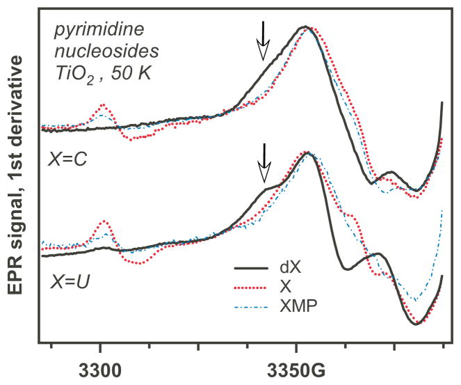
Figure 4.
EPR spectra from pyrimidine (X=C, U; Scheme 1) ribonucleosides (X, dotted lines), 2′-deoxyribonucleosides (dX, solid lines), and the corresponding ribonucleotide-5′-monophosphates (XMP, dash dot lines) on photoirradiated aqueous TiO2 at 50 K. The arrows indicate spectral regions where nucleotides and 2′-deoxynucleotides have different spectral features.

