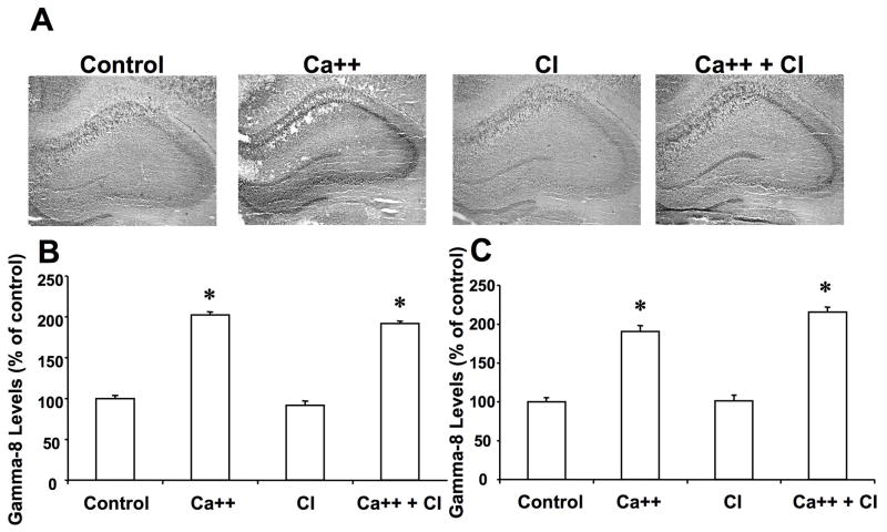
Figure 4. Effects of calcium and calpain inhibitor on γ-8 immunolabeling in hippocampus.
Thin frozen-thawed brain sections were incubated at room temperature for 90 min in the absence of calcium (Control), in the presence of 2.0 mM calcium (Ca++), and in the presence of 10 μM calpain inhibitor III (CI). They were then processed for immunohistochemistry with anti-γ-8 antibodies.
A. Representative images of brain sections incubated under each condition.
B & C. Quantitative analysis of images similar to those shown in A. Staining levels were analyzed in the cell bodies of field CA1 (B) and CA3 (C). Results were expressed as percent of the values found under control conditions and are means ± S.E.M. of 12 sections. *p<0.001.

