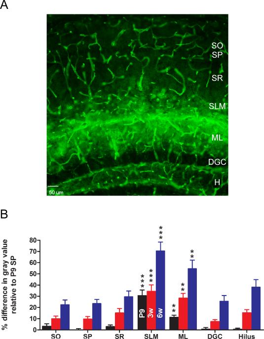
Figure 3. Laminar specificity of hippocampal AQP4 immunoreactivity.
A. AQP4 immunoreactivity from 6-week-old mouse hippocampus demonstrates laminar specificity. While blood vessels are labeled well in all laminae, parenchymal AQP4 immunoreactivity is strongest in CA1 stratum lacunosum moleculare (SLM) and dentate gyrus molecular layer (ML) and along the hippocampal fissure separating these laminae. Laminar boundaries are derived from Nissl co-labeling (not shown for clarity). Scale bar, 50 μm. SO, stratum oriens; SP, stratum pyramidale; SR, stratum radiatum; DGC, dentate granule cell layer; H, hilus.
B. Developmental and laminar-specific quantitation of hippocampal AQP4 immunoreactivity. Values are expressed as % difference in gray value compared with P9 stratum pyramidale (SP) (see Materials and Methods). P9, black bars; 3 weeks, red bars; 6 weeks, blue bars. Higher values indicate more intense immunoreactivity. **, p<0.01 and ***, p<0.001 compared with SP at same time point.

