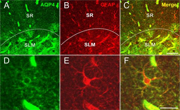
Figure 4. Colocalization of AQP4 and GFAP.
Immunoreactivity for AQP4 (A), GFAP (B) and merged (C, F) is shown at the hippocampal CA1 SLM/SR border (dotted line) in a 3-week-old mouse. Note that in the SLM, there is significant colocalization of GFAP and AQP4 on cells with the morphology of bona fide astrocytes, whereas in the SR, GFAP-labeled astrocytes are seen that are AQP4-negative. AQP4 labels blood vessels in both layers. Higher-power images (D-F) demonstrate AQP4 and GFAP colocalization on an astrocyte in CA1 SR from a 6-week-old mouse. Scale bars, 50 μm (top), 20 μm (bottom).

