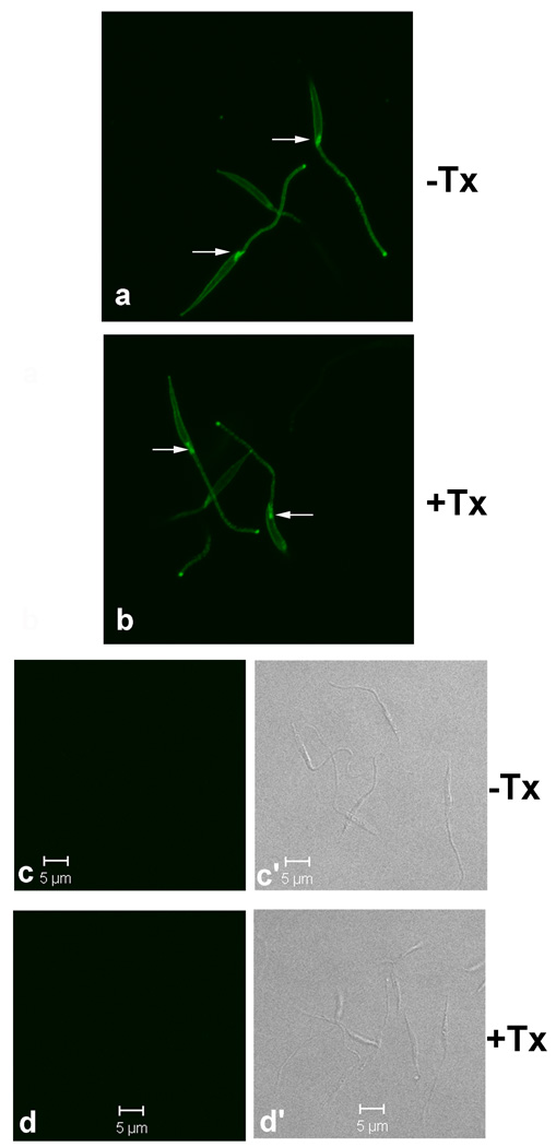
Figure 3.
Proteins in the flagellar pockets of metacyclic promastigotes are accessible to surface biotinylation. Metacyclic promastigotes were surface biotinylated in 1 mM Sulfo-NHS-biotin/HBSS (Panels a and b) or in HBSS alone (Panels c-c’ and d-d’), followed by fixation in 10% PBS-buffered formalin. Afterwards, the cells were incubated in 0.2% Triton X-100 (+Tx)/PBS or in PBS alone (−Tx). Cells were then incubated in FITC-conjugated extravidin and observed by confocal microscopy at 0.3 µm for z-section. Panels a and b shown stacked z-sections generated by the open access ImageJ software (rsbweb.nih.gov/ij/). Biotinylated flagellar pockets are marked by arrows. No background staining is observed from non-biotinylated cells (c and d) with DIC (c’ and d’) to show the cells.

