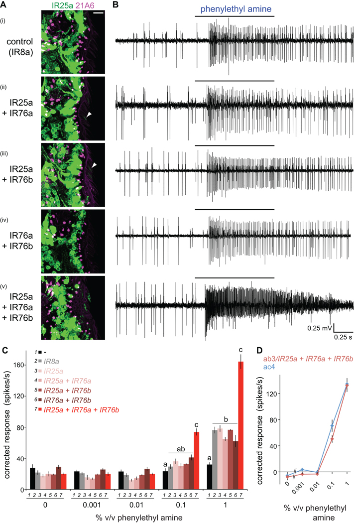
Figure 8. An olfactory receptor of three IR subunits.
(A) Immunostaining for IR25a (green) and the cilia base (21A6, magenta) in OR22a neurons expressing the indicated combinations of IRs. Weak cilia localization of IR25a in neurons co-expressing IR25a+IR76a or IR25a+IR76b is indicated by arrowheads. Genotypes: (i) UAS-IR8a/+;OR22a-GAL4/+, (ii) UAS-IR76a/UAS-IR25a; OR22a-GAL4/+, (iii) UAS-IR25a/+;UAS-IR76b/OR22a-GAL4, (iv) UAS-IR76a/+;UAS-IR76b/OR22a-GAL4, (v) UAS-IR76a/UAS-IR25a;UAS-IR76b/OR22a-GAL4.
(B) Representative traces of extracellular recordings of neuronal responses in OR22a neurons expressing the combinations of IRs shown to the left in (A) stimulated with phenylethyl amine (1% v/v). Bars above the traces mark stimulus time (1 s). Misexpression of a control receptor, IR8a, confers weak responsiveness to phenylethyl amine, which may reflect non-specific sensitization of these neurons to this odor, as IR8a does not localize to sensory cilia in the absence of IR84a (Figure 3A).
(C) Concentration-responses for phenylethyl amine in OR22a neurons expressing the combinations of IRs shown in the key. Genotypes are as in (A) as well as a no IR control (“-“) (OR22a-GAL4/OR22a-GAL4) and IR25a misexpression alone (UAS-IR25a/+;OR22a-GAL4/+). Mean responses are plotted (± s.e.m; n=12–28 sensilla; ≤4 sensilla/animal, male flies). For responses to 0.1% and 1% phenylethyl amine, bars labeled with different letters are significantly different (ANOVA p<0.0001).
(D) Comparison of concentration-responses for phenylethyl amine in OR22a neurons ectopically expressing IR25a+IR76a+IR76b (red) to those in endogenous ac4 sensilla (blue). Responses in OR22a neurons were corrected for background endogenous neural responses (black bars in (C)). Responses to phenylethyl amine in ac4 were measured in an IR8a1 mutant background to facilitate quantification of odor-evoked spikes in the absence of IR84a neuron activity (Figure 2B).

