
Fig. 1.
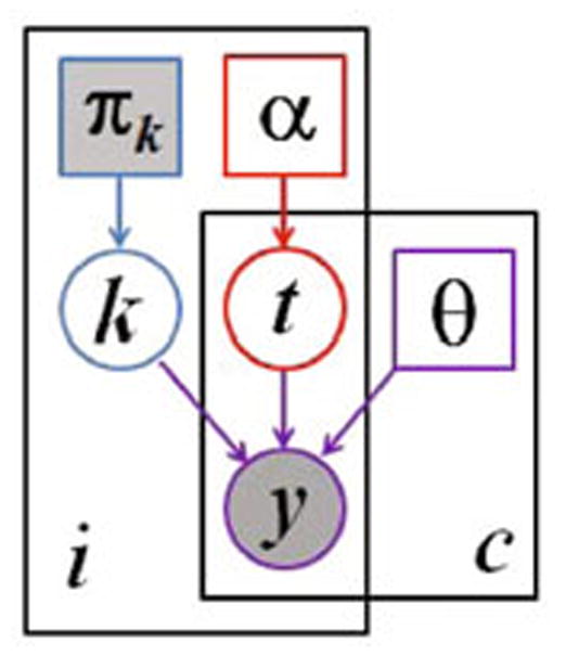
Graphical model for the proposed segmentation approach. Voxels are indexed with i, the channels are indexed with c. The known prior πk determines the label k of the normal, healthy tissue. The latent atlas α determines the channel-specific presence of tumor t. Normal state k, tumor state t, and intensity distribution parameters θ jointly determine the multi-modal image observations y. Observed (known) quantities are shaded. The tumor segmentation aims to estimate , along with the segmentation of healthy tissue p(ki|y).