Figure 5.
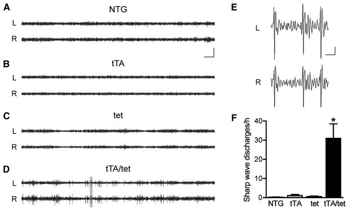
Epileptiform activity in parietal cortex of EC-APP mice. Bilateral EEG recordings were performed in 4–5-month-old mice of all four genotypes for 24 h. (A–C) Representative traces from control groups show normal EEG activity with no or very infrequent sharp wave discharges (SWDs). (D) In contrast, EC-APP (tTA/tet) mice displayed frequent SWDs. (E) The gray area in (D) was magnified to reveal the waveforms of the spike discharges in EC-APP mice. (F) Quantification of total SWDs per hour revealed a significantly greater frequency of SWDs in EC-APP mice than in all other groups. p<0.0005 for genotype by ANOVA and *p<0.05 vs. all other groups by Tukey post-hoc test. n=4–7 mice/group. L, left parietal cortex; R, right parietal cortex. Values are mean ± SEM. Scale bars: A, 10 s and 500 μV; E, 0.5 s and 100 μV.

