
Fig. 1.
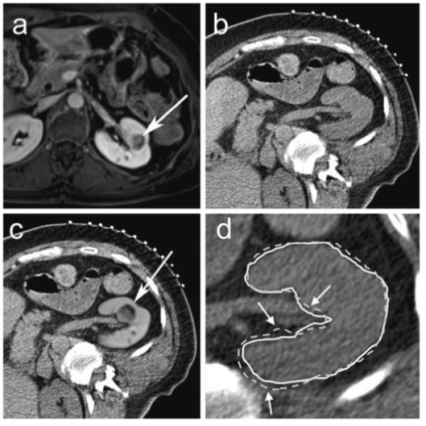
63-year-old female with left renal cell carcinoma. a Pre-procedural transverse contrast-enhanced spoiled gradient echo MR image of patient in supine position in a 1.5T scanner shows a 2.1 cm left renal mass (arrow). b Planning transverse unenhanced CT scan in the right posterior oblique position at the beginning of a CT-guided renal cryoablation shows the mass but the margins are not defined well. c MR image fused with CT image after non-rigid registration technique shows well-defined tumor in the left kidney (arrow). d The renal contour derived from registered MR images using non-rigid registration (solid white line) matches the renal contour on the CT image (white arrows) better than the renal contour derived from registered MR images using rigid registration (dotted white line)