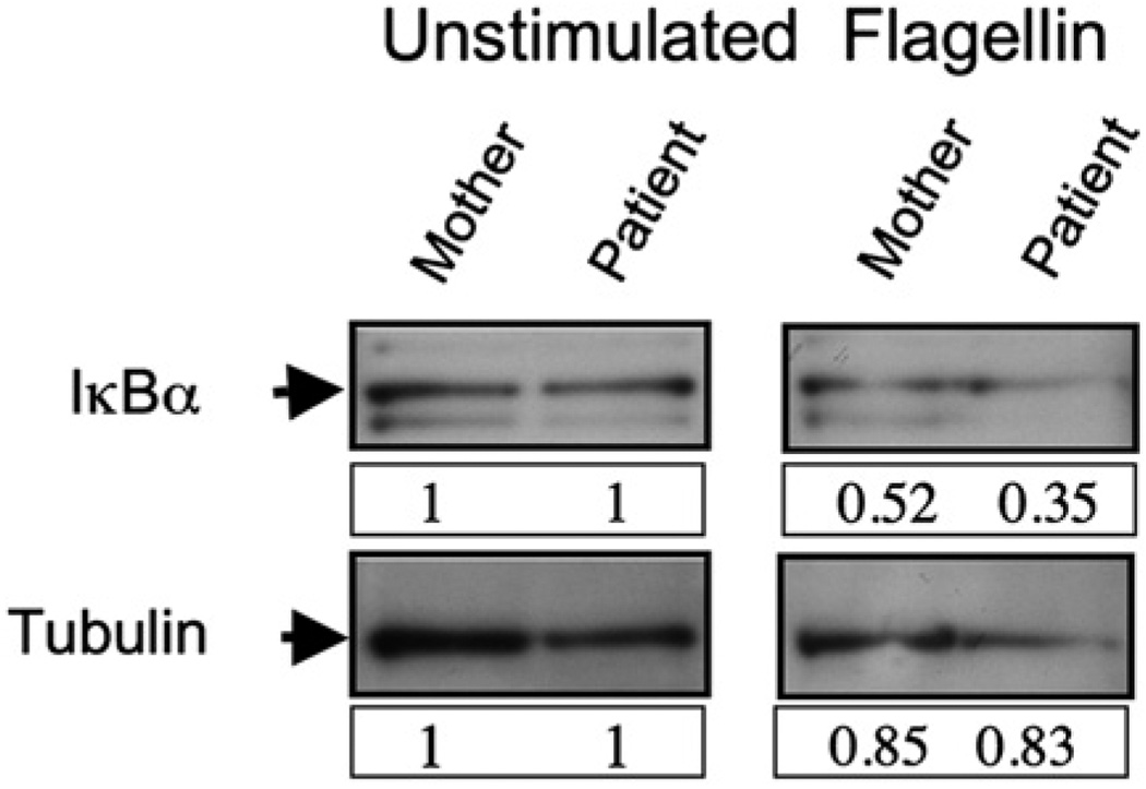
FIG 5.
Normal IκB degradation after exposure of patient cells to the TLR5 ligand flagellin. Whole-cell lysate IκBα Western blot (top) of unstimulated (left) or TLR5 ligand-stimulated (right) PBMCs. Membranes were stripped and reprobed for tubulin (bottom) as a loading control. Lysates from maternal PBMCs (left lanes) were compared with patient PBMCs (right lanes). Numbers depict band intensity relative to control, and blots are representative of 3 independent experiments.

