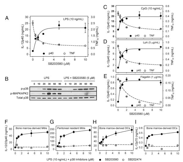
FIGURE 1.
MAPK/p38 inhibition increases IL-12 production by stimulated macrophages and DCs. A, Macrophages (3 × 10⁵ cells) were pretreated with increasing concentrations of SB203580 for 1 h and then stimulated with LPS (10 ng/ml) overnight. Supernatants were harvested, and IL-12p40 and TNF-α production were determined by ELISA. B, Macrophages (2 × 10⁶ cells) were pretreated with SB203580 (5 μM) for 1 h. Cells were then stimulated with LPS (10 ng/ml) for 0, 10, 20, 30, 60, and 90 min. Cell lysates were prepared for Western blot analysis to detect p-p38, p-MAPKAPK2, and total p38 protein. C–E, Macrophages (3 × 10⁵ cells) were pretreated with increasing concentrations of SB203580 for 1 h and stimulated with CpG (10 ng/ml) (C), lipoprotein A(5 μg/ml) (D), or flagellin (1 μg/ml) (E) overnight. IL-12p40 and TNF-α production were determined as described above. F–I, BMMφs (F), peritoneal macrophages (G), and BMDCs (H, I) were pretreated with increasing concentrations of SB203580 (●), or inactive SB202474 (엯) for 1 h and then stimulated with LPS (10 ng/ml) overnight. Supernatants were harvested for ELISA analysis of IL-12p40 (F–H) or IL-12p70 production (I). Values represent the mean ± SD of triplicate determinations, and the data represent one of three independent experiments.

