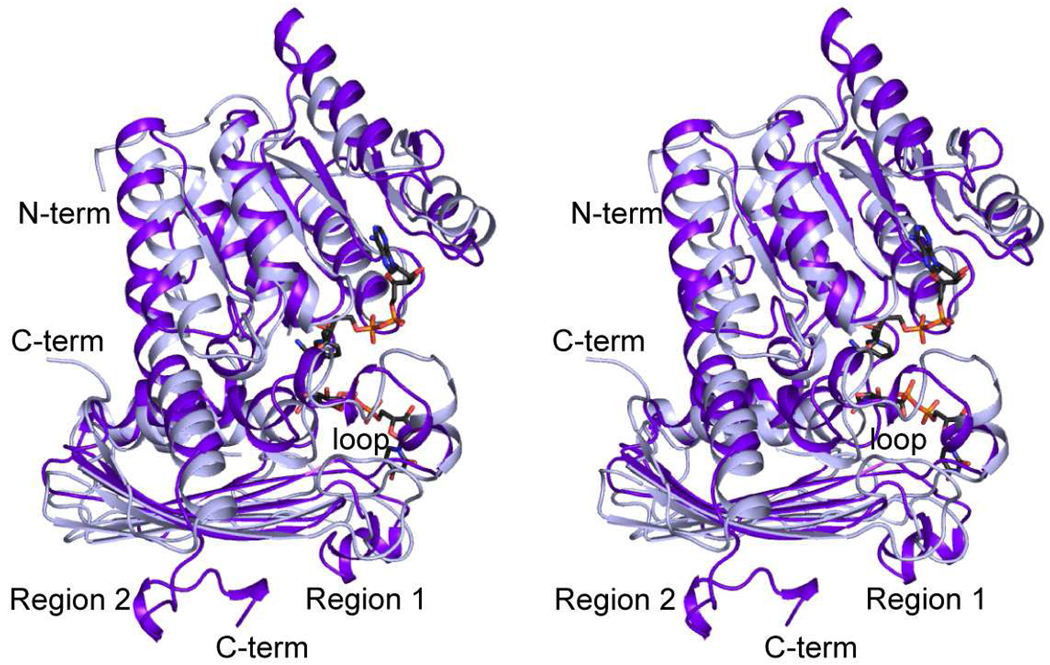
Figure 4.
Superposition of the ribbon representations for the P. aeruginosa and C. violaceum enzymes. The P. aeruginosa and C. violaceum WlbAs are highlighted in purple and light blue, respectively. X-ray coordinates for the P. aeruginosa enzyme were determined in this laboratory and deposited in the Protein Data Bank (accession no. 3OA0).

