Figure 2.
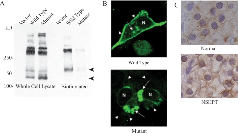
Cellular location of CaSR. A, Western blotting of the CaSR in whole cell lysates (left) and biotinylated cell surface proteins (right) from HEK293 cells transfected with vector and cDNA expression vectors for WT and mutant CaSR. The bottom two bands, corresponding to 140 and 160 kD, are the monomeric forms of CaSR; the upper bands are multimers or aggregates of the protein under reducing conditions. B, Dual fluorescence confocal microscopy. HEK293 cells were transfected with vectors expressing WT (top panel) or mutant (bottom panel) CaSR. Most WT CaSR is targeted to the cell surface (arrowheads), with little remaining intracellularly (arrows), whereas most of the mutant CaSR remains intracellular. C, Immunohistochemical staining of CaSR in parathyroid tissue. Top, Normal adult control shows anti-CaSR staining mainly along the surface of the chief cells; bottom, parathyroid tissue from the index patient shows perinuclear staining with absence of cell surface staining.

