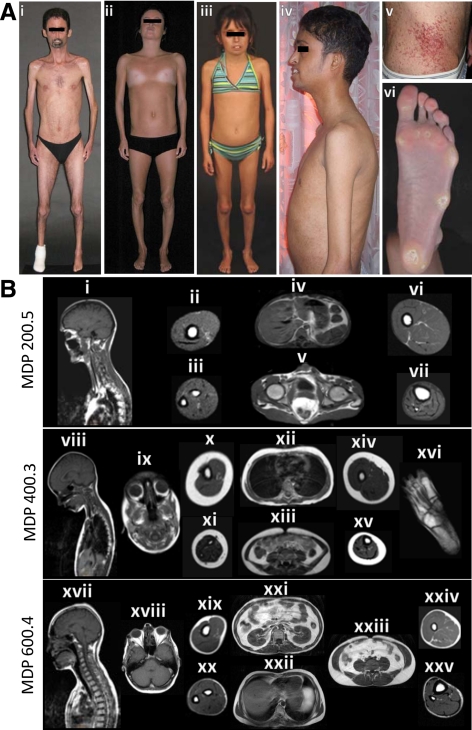
Figure 1.
MDP patients and their body fat distribution pattern on MRI. A, i, MDP 200.5, anterior view of a 36-yr-old male showing generalized lipodystrophy, reduced muscle mass, and prominent maxillary incisors. He had pinched nose, small mouth, and contractures of the elbows. There was no loss of scalp hair and he had graying of hair on the chin. ii, MDP 300.4, anterior view of a 19-yr-old Italian female showing generalized lipodystrophy and lack of breast development. She had pinched nose and small mouth. iii, MDP 400.3, anterior view of a 10-yr-old French Canadian girl with small mouth, pinched nose, and bilateral coxa valga. Her body fat distribution was normal. iv, MDP 500.4, lateral view of a 25-yr-old Indian male showing generalized lipodystrophy, muscle wasting, and prominent maxillary incisors. v, Telangiectasias on the abdomen of MDP 200.5. vi, Marked loss of sc fat from the sole and calluses in the patient MDP 200.5. B, Sagittal T-1-weighted MRIs of the head, neck, and thorax through midline or orbit and axial MRIs of the chest, abdomen, arm, forearm, thigh, calf, and foot from various MDP patients. MRIs of MDP 200.5 show near total loss of sc fat from the head, neck, and thorax (i), arm (ii), forearm (iii), abdomen (iv and v), thigh (vi), and calf (vii). Retroorbital, ip, and bone marrow fat depots were well preserved. MRIs of MDP 400.3 show normal amounts of sc fat in the head and neck (viii and ix), and thorax (xii), normal retroorbital (ix), ip (xiii), retroperitoneal, and bone marrow fat as well as in the arm (x), forearm (xi), thigh (xiv), and calf (xv). Plantar fat (xvi) was reduced. MRIs of MDP 600.4 show moderate loss of sc fat in all the areas including the head and neck (xvii), thorax (xxii), arm (xix), forearm (xx), thigh (xxiv), and calf (xxv). Retroorbital (xviii), intraabdominal, and bone marrow fat depots were well preserved. A significant amount of intraabdominal (xxi and xxiii) fat was noted.

