
Figure 2.
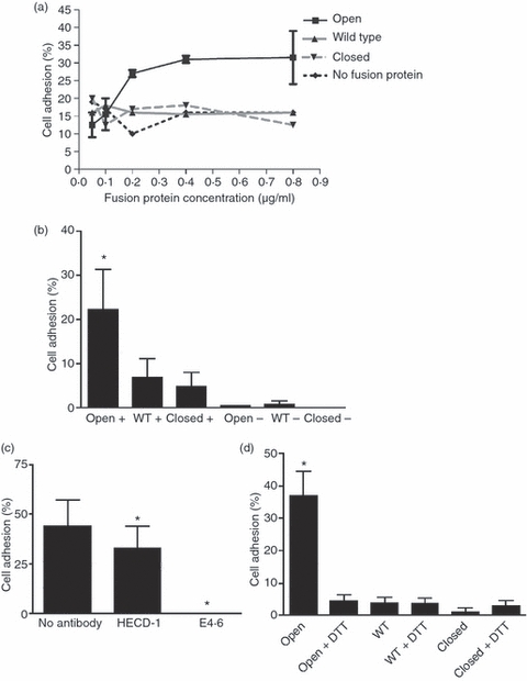
Adhesion of MCF-7 cells to human αE I domain fusion proteins. (a) The percentage binding of MCF-7 cells to serial dilutions of fusion protein in the presence of Mn2+. Bovine serum albumin (BSA) (1%) was used as a negative control. (b) The percentage of MCF-7 cell binding to ‘open’, wild-type and ‘closed’ fusion proteins in the presence (+) and absence (−) of Mn2+. Background adhesion to BSA was subtracted. Combined results from three separate experiments are shown, and error bars represent the standard error of the mean (SEM); *, P < 0·05. (c) The effect of mAbs (HECD-1 and E4.6) to E-cadherin on MCF-7 cell binding to the ‘open’ fusion protein in the presence of Mn2+. Background adhesion to BSA was subtracted. Combined results from three separate experiments are shown, and error bars represent the SEM; *, P < 0·05. (d) Percentage of MCF-7 cell binding to ‘open’, wild-type and ‘closed’ fusion proteins in the presence and absence of dithiothreitol (DTT). Background adhesion to BSA was subtracted. Combined results from three separate experiments are shown, and error bars represent the SEM; *, P < 0·001. WT, wild type.