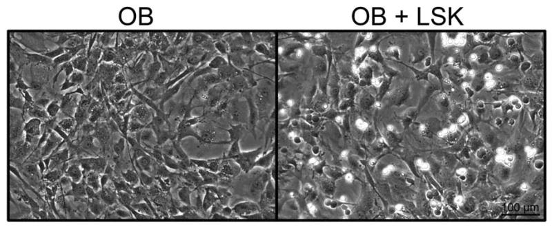
Figure 1.
Micrographs of OB cultured for 4 days in the presence or absence of LSK cells and cytokines. No significant differences were observed in OB morphology or confluence when cultured with LSK cells and cytokines. The hematopoietic cells are the smaller, more spherical cells which refract the light differently than the OB and appear more white in the image.

