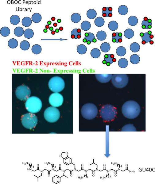
Figure 5.

A screening protocol for the identification of highly selective ligands for an integral membrane protein [17]••. See text for details. The fluorescent micrographs show the results from a screen employing cells that do (red-labeled) or do not (green-labeled) express VEGFR2. When an OBOC library of peptoids containing about 260,000 compounds was employed, several thousand beads displaying bound red and green cells were observed (left micrograph). Only five beads that bound almost red cells almost exclusively were observed. The structure of the peptoid, GU40C, displayed by the bead pictured is shown.
