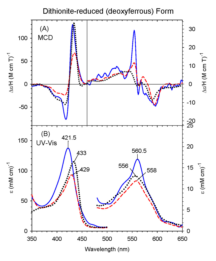
Figure 3.
MCD (A) and electronic (UV-Vis) absorption (B) spectra of deoxyferrous ΔC436S CYP2B4 mutant (solid line), human H93Y Mb (dashed line) and SW Mb (dotted line). The spectra for the H93Y Mb and SW Mb were obtained in 0.1 M potassium phosphate (pH 7.0) at 4 °C and for the ΔC436S CYP2B4 mutant in 70% (v/v) glycerol and 30% 0.3 M potassium phosphate (pH 8.0) at −50 °C.

