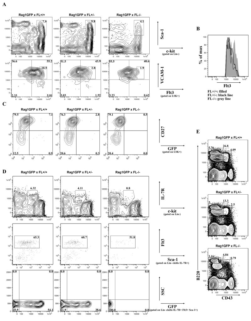
Figure 2.
FL haploinsufficiency reduces LHP and RAG1 locus activation. (A) Flow cytometric analysis of LSK+ BM cells from RAG1-GFP x FL+/+, RAG1-GFP x FL+/−, and RAG1-GFP x FL−/− mice (top panels). Differential expression of Flt3 and VCAM-1 in LSK+ BM cells to discriminate HSC/MPP, GMLP, and LMPP (bottom panels). Boxed regions indicate Flt3hi GMLP. (B) Histogram of boxed region from (A) bottom panels, indicative of Flt3 expression in Flt3hi GMLP in different FL genotypes. (C) Flow cytometric analysis of LSK+ BM cells from RAG1-GFP+ x FL mice to distinguish HSC, MPP, and LHP. A littermate GFP- control was analyzed in each experiment to determine GFP+ gates (data not shown). (D) Flow cytometric analysis of Lin- BM cells to distinguish CLP: Lin- c-kitlo IL-7R+ (Top panels). CLP were further discriminated by Flt3 and Sca-1 expression (middle panels). GFP expression within Lin- c-kitlo IL-7R+ Flt3+ Sca-1+ CLP (bottom panels). GFP+ gates were determined by analysis of Lin- c-kitlo IL-7R- Flt3+ cells which do not express GFP (data not shown). (E) Flow cytometric analysis of BCP. Pre-Pro-B/Pro-B cells are B220+ CD43+, Pre-B cells are B220+ CD43-, and naïve/mature B cells are B220hi CD43-. Data are representative of ≥ 5 mice/genotype and ≥ 3 independent experiments.

