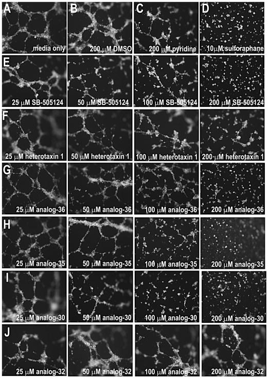
Figure 7. Heterotaxin inhibits angiogenesis in human cells.
Human umbilical vein endothelial cells (HUVECs) form tubes when cultured in the presence of media alone (A), DMSO (B), unsubstituted pyridine (C), or the inactive heterotaxin analog 32 (J). In contrast, HUVECs cultured in the presence of a known anti-angiogenic agent (D, sulforophane; Bertl et al., 2006) are unable to form tubes. Although the original heterotaxin molecule 1 (F) exhibits only very mild anti-angiogenic effects in this assay (at 6 hr), the other active heterotaxin analogs 36 (G), 35 (H) and 30 (I) have obvious concentration-dependent anti-angiogenic properties comparable to a known TGF-β signaling inhibitor (E, SB-505124). Cells were stained with Calcein AM to confirm viability. (See Figure S3 for effect of analog 30 on tumor cell growth.)

