Figure 1.

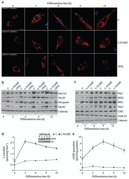
NO regulates myogenesis and mitochondrial network formation through cGMP. Myogenic precursor cells transfected with the red fluorescent mitochondrial protein mitoDsRed were treated with l-NAME, ODQ or vehicle (C) and differentiated by serum withdrawal for up to 12 h. (a) Mitochondrial morphology detected by transient transfection with mitoDsRed; nuclei are stained for Myo-D (blue) to distinguish myogenic cells from possible contaminating cells. Bar: 10 μm. (b) Expression of the differentiation markers Mef-2A, Myo-D, myogenin and sarcomeric myosin (MyHC) as detected by western blot analysis. (c) Expression of the mitochondrial proteins mitofusins (Mfn)-1 and 2, Opa1, Drp1, cytochrome c (CytC) and cytochrome c oxidase subunit-IV (COX-IV). Calnexin (Clx) was used as a loading control in the experiments in panels b and c. (d) NOS activity measured as the conversion of l-[3H]-arginine into l-[3H]-citrulline. The inset shows the levels of expression of nNOS during myogenic differentiation. Clx was used as a loading control. (e) Generation of cGMP. The images are from one of four independent reproducible experiments; the graphs represent the values±S.E.M. (n = 4)
