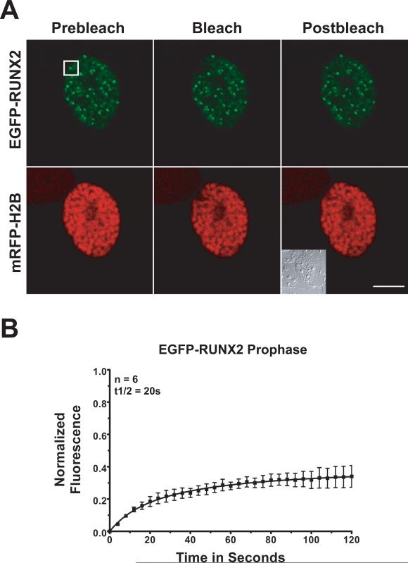
Fig. 3. Photobleach recovery of RUNX2 on chromosomes during prophase.
Multiple prophase cells expressing EGFP-RUNX2 and mRFP-Histone H2B were used for FRAP analysis. (A) The images show the region of interest in cells before photobleaching, just after photobleaching, and 120 seconds later when the fluorescence had recovered. The size bar is 10 μm. (B) Normalized mean fluorescence recovery curves over time for 6 cells were calculated for chromosome bound RUNX2; the means were plotted with standard errors for each time point. The kinetics of recovery measure the interaction of the protein with its binding site. Cells in prophase show a half-time of recovery (t1/2) of 20 seconds. The large immobile fraction (67%) represents RUNX2 in complexes that are tightly bound and do not dissociate over the time course of the experiment.

