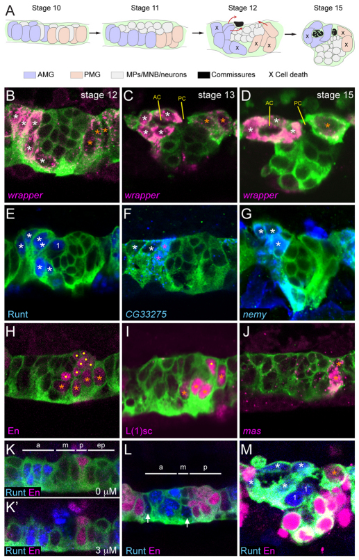
Fig. 1.
Anterior midline glia (AMG) and posterior midline glia (PMG) differ in gene expression and origins. (A) Schematic summary of midline glia (MG) positioning and migration in sagittal views. The schematic depicts an idealized view; actual segments vary in MG number and position. Colored objects represent nuclei, and pathways of AMG and PMG migration are indicated at stage 12 by red arrows. MNB, median neuroblast; MP, midline precursor. (B-M) Fluorescence confocal images of single segments in sagittal view from sim-Gal4 UAS-tau-GFP embryos. Anterior is to the left and dorsal (internal) is up. White asterisks indicate AMG; orange asterisks, PMG; dots, midline neurons; ‘1’, MP1 neurons. RNA (italicized) and antibody (non-italicized) stains and corresponding colors are indicated in the lower left corner of each panel except for anti-GFP staining (green) that is present in all images. (B) During stage 12, AMG (high levels of wrapper RNA) and PMG (low levels of wrapper RNA) are elongating and moving to the dorsal (internal-most) surface of the CNS. Midline neurons are the centrally located wrapper− cells flanked by wrapper+ MG. (C) During stage 13, AMG migrate posteriorly above and below the anterior commissure (AC). PMG abut the posterior commissure (PC). (D) By stage 15, AMG completely surround the AC and are poised to ensheath the PC. Most PMG have undergone apoptosis; the remainder stay in contact with the PC. (E-G) Runt is present in all AMG and the MP1 neurons, whereas CG33275 and nemy expression is restricted to AMG only. The AMG closest to the midline neurons and developing commissure (pink asterisks) have high levels of CG33275, compared with those that are more distant (white asterisks). (H-J) En and L(1)sc are present in all PMG and in a subset of midline neurons whereas mas is expressed in only one or two PMG in each segment. (K,K′) Two focal planes (separated by 3 μm) of a stage 10 segment; Runt is present in four to five cells in the anterior cells (a), Runt and En are absent from the middle (m) and extreme posterior cells (ep), and En is present in two cells in the posterior (p). (L) During stages 10-11, Runt expands to additional MG (arrows) and the number of En+ cells increases (stage 11 is shown). The segment is divided into anterior (a) runt+ en−, middle (m) runt− en− and posterior (p) runt− en+ domains. (M) In stage 15 MG, Runt and En are present in AMG and PMG, respectively.

