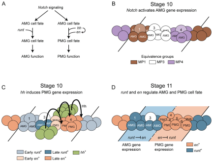
Fig. 8.
Model of hh, en and runt regulation of midline glia (MG) cell fate. (A) Flow diagram describing the genetic regulation of anterior midline glia (AMG) and posterior midline glia (PMG) development. (B,C) Schematics of stage 10 segments. Anterior is to the left and lines indicate segmental boundaries. (B) Notch signaling results in the activation of MG gene expression and the formation of MP1 (1), MP3 (3) and MP4 (4) from three equivalence groups. (C) Hh, originating in cells lateral to the midline, signals (arrows) to the midline cells that are posterior to the early en+ cells, activating late en expression. Late runt is initiated in cells flanking the early runt+ cells. MP1 is shown arising from early runt+ cells, as it is runt+ and forms during stage 10 prior to the initiation of late runt expression. The early en+ cells are shown as PMG and not AMG, MP1, MP3-6, or the median neuroblast (MNB), because: (1) AMG, MP1 and MP3 arise from en− cells (Wheeler et al., 2006); (2) MP4 arises from en− cells just posterior to the early en+ cells (Fig. 2B′); and (2) MP5, MP6 and the MNB all arise posterior to MP4 (Wheeler et al., 2006). This leaves only PMG as the descendants of the early en+ cells. The other two PMG arise from the late en+ cells, along with MP4-6 and the MNB. (D) Schematic of early stage 11, in which the MP4 has divided into two ventral unpaired median 4 (VUM4) neurons (4). Other MPs are designated 1, 3, 5 and 6, along with the MNB. Dashed line indicates the boundary between runt+ and en+ domains. In the runt+ domain, runt represses en expression, and in the en+ domain, en represses runt expression. This cross-repression maintains distinct AMG and PMG cell fates. MP, midline precursor.

