Fig. 3.
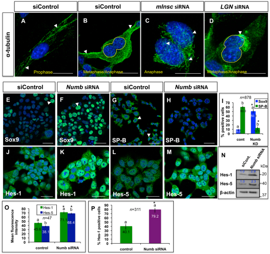
Functions of polarity proteins in lung epithelium in vitro. (A,B) Immunocytochemistry with α-tubulin antibody shows well-organized and oriented spindle fibers (arrowheads) in MLE15 cells during early (A) and late (B) mitosis. (C,D) Spindle fibers are disorganized/disoriented (arrowheads) in mitotic MLE15 cells after Insc or Gpsm2 knockdown. (E-M) Immunocytochemistry shows that MLE15-positive cells (arrowheads) for Sox9 (F), Hes-1 (K) or Hes-5 (M) increase with strong nuclear staining, while SP-B-positive cells (H) decrease after Numb knockdown. (I) Quantitation of Sox9- or SP-B-positive cells, which is expressed as a percentage of all counted MLE15 cells, of the experiments shown in E-H. Bars carrying the same letter (a,b) in I, O or P are significantly different from one another (*P<0.05; Student's t-test). Data are mean±s.e.m. (N) Western blot of the experiments shown in J-M. (O) Means fluorescence intensity of Hes-1 or Hes-5 staining for experiments showing in J-M. (P) Quantitation of Hes-1-positive cells, which is expressed as a percentage of all counted MLE15 cells, of the experiments shown in J-K. In O,P, Error bars indicate s.e.m. Scale bars: 50 μm.

