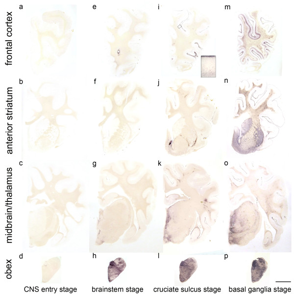
Figure 2.
Classification of the PrPSc spread during disease development in classical scrapie.The examined classical scrapie cases classified into four stages of PrP spread according to certain affected neuroanatomical sites (PET blots, mAb P4). In the CNS entry stage (a - d) only discrete PrPSc deposits are visible in the obex region, while in the brainstem stage (e - h) PrPSc aggregates are clearly visible in the brainstem and start to appear in more rostral structures. Once PrPSc deposits can be found in the deep cortical layers of the frontal cortex (i), the cruciate sulcus stage (i - l) is reached. In the basal ganglia stage, intense deposits in basal ganglia and thalamic nuclei can be found (m - p). Brain sections shown for the first, third and fourth stage derived from sheep with the genotype ARQ/ARQ while the sheep whose brain sections are depicted in the brainstem stage carried the genotype ARH/VRQ (bar = 5 mm).

