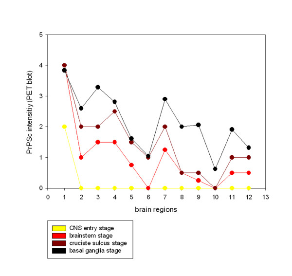
Figure 4.
Accumulation of PrPSc in different brain regions during disease progression: The four stages of the examined classical scrapie cases are depicted in four overlying graphs that illustrate how PrPSc aggregates (PET blot method, mAb P4) are increasingly accumulated in the brains from caudal (left) to rostral (right). Evaluation of PrPSc intensity was performed on a scale from 0.5 - 4 (see material and methods) and shown for the following brain areas: 1 dorsal motor nucleus of the vagus nerve (DMNV), 2 inferior olive, 3 dorsal tegmental nucleus, 4 cerebellar molecular layer, 5 cerebellar granular layer, 6 cerebral peduncle, 7 central grey (mesencephalon), 8 caudate nucleus, 9 ventral pallidum, 10 rostral commissure, 11 cruciate sulcus, 12 frontal white matter.

