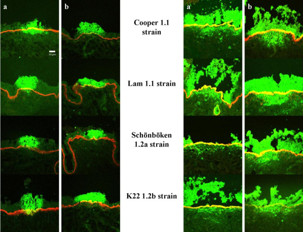
Figure 5.
Confocal fluorescent images of bovine respiratory (a) and genital (b) mucosa explants inoculated with different BoHV-1 subtypes at 24 h pi (left side) and 72 h pi (right side). Viral antigen is colored with an FITC®- labeled goat anti-IBR polyclonal antiserum. Collagen VII is marked with mouse anti-collagen VII and goat anti-mouse Texas Red® antibodies.

