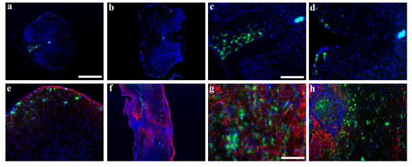
Figure 3.
5 weeks post SCI, CXCR4 is found in 3 different areas of the spinal cord. Thoracic cross sections demonstrate CXCR4-GFP in the ependymal layer (a-d), peripherally toward the meninges (a-e) and in the dorsal funiculus rostral to the injury (a, c) but not caudal to it (b, d)(green:CXCR4-EGFP, blue:Hoechst). Longitudinal sections show CXCR4-GFP+ cells in the lesion epicenter as noted in f and in the cavitation in h, but also outside the lesion (f, h). Some CXCR4-GFP cells were found within the GFAP+ scar, but were distinct from the GFAP+ cells (g)(red:GFAP, green:CXCR4-EGFP, blue:Hoechst). Scale bars: a-b, f: 200 μm, c-d, e, h: 50 μm, g:25 μm

