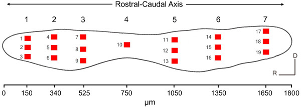
Figure 1.
Pre-selected locations on the goldfish saccule for hair cell quantification. Schematic drawing of a left saccular epithelium from a goldfish, showing the 19 regions (50 μm × 50 μm each) in which phalloidin-labeled hair cell bundles were counted. Each column of boxes was represented as a rostral-caudal axis number from 1 to 7, with 1 and 7 being the most rostral and caudal, respectively. The corresponding distance (μm) from the rostral tip of the saccule for each rostral-caudal axis number is marked on the scale below the figure. Each row of boxes was represented as a dorsal-ventral axis number from 1 to 3, with 1 being dorsal and 3 being ventral.

