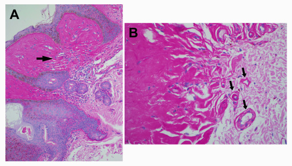
Figure 2.
Papillary dermis. (a) Deposition of PAS positive and diastase-resistant material from Patient II-1 of Family 1. Rare inflammatory cells are seen in the upper dermis (original magnification X 200). (b) PAS positive material deposited around blood vessels which have thickened and hyalinized walls. (arrows, original magnification X 400). There was no evidence of infarction.

