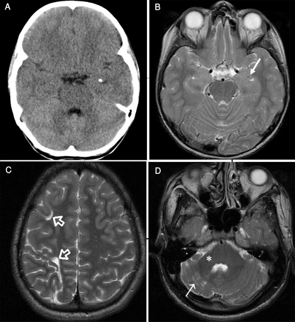
Figure 3.
Brain CT and MRI of Patients 1 and 2. (a) Brain CT image of Patient II-6 of Family 1 at age of 10 years showing a small calcification in the left amygdale. Calcification in the right amygdale was seen on another section (not shown). (b) Brain axial T2-weighted MR image of the same patientat the level of temporal lobes showing a tiny hypointensity (arrow) representing amygdala calcification. The mesial temporal lobes were otherwise normal. (c) Brain axial T2-weighted MR images of Patient II-1 of Family 1 showing enlarged sulci and small gyri in the watershed zones of the right frontal lobe (open arrows) associated with loss of white matter due to old ischemia. (d) The right cerebellar hemisphere of the same patientwas small with widened folia, white matter gliosis (arrow), and a small right middle cerebellar peduncle (asterisk). Remodeling of the posterior fossa confirmed the long-standing nature of this abnormality. CT and MRI scans on patients from Families 2 and 3 were normal.

