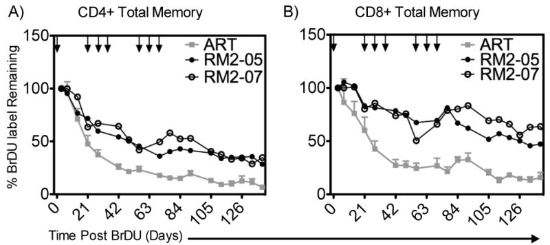
Figure 7. %BrDU Label Retained in Total Memory CD4+ T-cells in the Blood.

BrDU was used to label cells dividing in response to rsIL-7gly administration following the first of nine doses. The percentage of BrDU+ cell immediately post labeling was set to 100% to normalize the rate of decline in all the macaques. Where applicable rsIL-7gly administration is indicated [↓]. (A) The percentage of BrDU label retained in CD4+ memory cells in the peripheral blood (B) The percentage of BrDU label retained in CD8+ memory cells in the peripheral blood. ART controls (mean +- SEM) (grey square); RN2-05 (closed circle) and RM2-07 (open circle).
