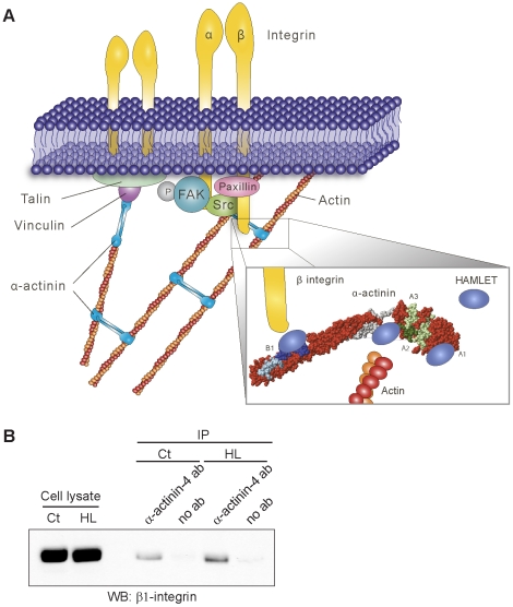
Figure 6. Schematic model of HAMLET-α-actinin interactions.
A) The figure is based on information available in MolMol (PDB codes 1SJJ and 1HML) and on results from the peptide-binding assay and not on actual three-dimensional structural data of HAMLET bound to α-actinin-4. α-Actinin forms a bridge between the cytoskeleton and the integrins through the actin- and β integrin-binding sites but also through vinculin and talin. By binding to α-actinin, HAMLET may disrupt focal adhesions (enlargement, lower right). HAMLET interacts with the actin-binding domain of α-actinin and fits into the pocket formed between the actin-binding domain and the first spectrin repeat. In addition, HAMLET binds adjacent to the β integrin-binding site. B) Cell extracts from untreated or HAMLET-treated cells were used for co-immunoprecipitation with anti-α-actinin-4 antibodies. Blots stained with anti-β1 integrin antibodies show that α-actinin-4 and β1 integrin still interact with each other in the presence of HAMLET.

