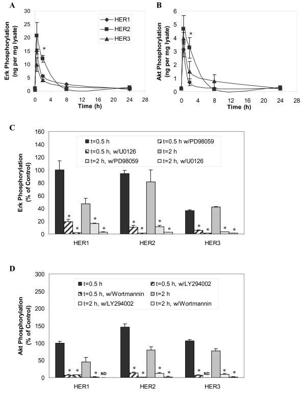
Figure 4.
Erk and Akt activation in response to EGF stimulation in HMEC. Phosphorylation levels of A) Erk and B) Akt were examined in HMEC parental HER1 cell (diamonds), HER2 (squares) and HER3 (triangles) using a commercial 96-well-plate ELISA. Lysate sample were collected from cells treated with 12 ng/ml EGF for 0, 0.5, 2, 8 and 24 h. Results are presented as ng pErk or pAkt per mg lysate protein. Phosphorylation level of C) Erk and D) Akt were also examined when cells were pretreated with MAPK inhibitors (25 µM PD98059 and 10 µM U0126) and PI3K inhibitors (20 µM LY94002 and 200 nM wortmannin). Data points (or columns) and cross bars represent the average and standard deviation, respectively, of two biological replicates, each from a separate culture dish. In panels A and B, the symbols denote statistically significant differences between the parental HER1 and HER2 (*) or HER3 (#) by t-test. In panels C and D, results were normalized based on the phosphorylation level of the control sample (0.1% DMSO alone) from the parental HER1 cells after 0.5 h EGF stimulation. In the presence of wortmannin, Akt phosphorylation levels of parental HER1 cell line at t = 2 hr and HER3 cell line at t = 0.5 hr were below assay detection limit. The asterisks in panel C and D denote statistically significant differences between inhibitor-treated sample and its control (0.1% DMSO in absence of any inhibitor) in each individual cell line at every time point.

